Protocol: Immunofluorescence on chemically fixed cells
On-chip chemical fixation and immunofluorescence of HER2 receptor of breast cancer cell membrane
Materials
- Cell medium
- 1X PBS (also used as mounting medium)
- Blocking buffer: NH4Cl 50 mM; BSA 0.5% in PBS
- BB700 Mouse Anti-Human Her2/Neu (anti-HER2 Ab, 1:100 dilution in blocking buffer)
- Mineral oil for cell culture
- Stereomicroscope
Cell culture on VersaLive
Follow the protocol relative to the cell culture on VersaLive.
On-chip chemical fixation
- Empty all ports
- PBS washing (1x), 20 μL in all ports
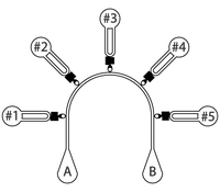
- 10 min perfusion of PFA 4% in ports #1 to #5 (leave ports A and B empty)
- PBS washing (2x), 20 μL in all ports
- 5 min perfusion PBS washing, 20 μL in ports #1 to #5 (leave ports A and B empty)
- 30 min perfusion blocking buffer, 20 μL in ports #1 to #5 (leave ports A and B empty)
- Remove blocking buffer, fill all ports with 20 μL PBS
- [Optional pause point] Add 2.5 μL mineral oil in all ports to prevent evaporation, store at 4˚C
On-chip immunostaining
- 10 min perfusion anti-HER2 Ab, 20 μL in ports #1 to #5 (leave ports A and B empty)
- PBS washing (2x), 20 μL in all ports
- Mounting medium: 20 μL PBS in all ports + 2.5 μL mineral oil to prevent evaporation
- Store at 4˚C

